
By Randy Griffith
The Tribune-Democrat, Johnstown, Pa.
WINDBER
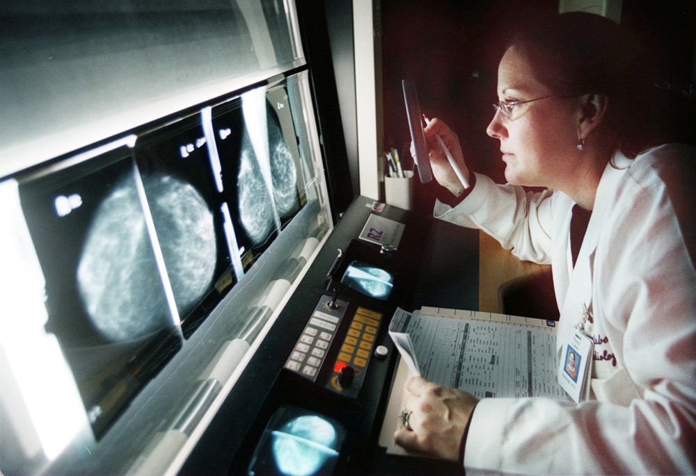
The region’s first automated whole breast ultrasound helps Windber Medical Center and its Joyce Murtha Breast Care Center remain a leader in the detection and treatment of cancer and other diseases of the breast, leaders say.
The breast volume scanner has been shown to detect twice as many cancer lesions than mammography alone in women whose breast tissue is dense.
“Cancer is harder to see in the dense breasts,” said Cindy Cwik-Esposito, radiology director. “We thought this would be a good thing to utilize for our patients with dense breasts. It will give you a better diagnosis.”
Earlier this year, Pennsylvania joined a growing number of states requiring mammography providers to notify women categorized as having dense breast tissue about their condition.
The laws stem from a 2009 incident in which a Connecticut woman with dense breast tissue was diagnosed with advanced breast cancer that was missed on her mammogram. She told state legislators she had not been informed by her doctor that she had dense breast tissue.
The whole breast ultrasound creates a 3-D image of the entire breast. This has been shown not only to find more cancer, but it helps surgeons and interventional radiologists plan treatment for known tumors.
Automated breast volume scanners reduce the chance of operator error because they use robotics to move across patients’ skin, taking thousands of images that are combined digitally into a 3-D model.
Mammography remains the gold standard for early diagnosis of breast cancer, but whole breast ultrasound will provide an additional view for select patients.
Doctors are already using magnetic resonance imaging as a second look in some women.