
By Delthia Ricks
Newsday.
MELVILLE, N.Y.
The human brain, a 3-pound organ and source of symphonies, mathematics, politics, hate and crime, is often hailed as the most complex biological entity in the known universe.
Some scientists call it a universe itself, one of the last great frontiers.
In recent months, scientists have uncovered some of the brain’s deepest secrets. One team has revealed how sleeping posture is important to brain health. Another has elucidated the circuitry that is cued when confronted with something unexpected, how the brain reacts to the element of surprise. Both are providing new clues about the life’s master organ.
The discoveries arrive only two years into a grand challenge by the Obama administration, which has called on neuroscientists to map the human brain. Most investigators had been racing all along, some searching for the switch that turns on dreams; others on the hunt for the causes of neurodegenerative disorders. One goal is to further demystify the organ’s bewildering circuitry, characterized by 100 billion connections among brain cells.
Biophysicist Partha Mitra of Cold Spring Harbor Laboratory, a 2014 recipient of a Brain Initiative grant, said despite centuries of cumulative knowledge, more is known about some of the deepest reaches of the cosmos than regions within the labyrinths of the brain.
“I have said that many, many times,” noted Mitra, who is using his $300,000 grant to develop “conceptual and physical” tools to help neuroscientists better explore the brain. He is also leading the lab’s Mouse Brain Architecture Project, an ambitious effort to decipher how neurons, brain cells that underlie all higher function, connect.
In the human brain, he said, a single neuron through its branching projections can make up to 10,000 connections, contributing to the billions upon billions brainwide.
Yet, Mitra isn’t alone in his pursuit to peel away some of the brain’s mystery.
Scientists at Stony Brook University’s School of Medicine are asking a deceptively simple question: Does a lateral sleeping position promote brain health? So far, their investigative evidence suggests a resounding yes.
Dr. Helene Benveniste and her team have found that sleeping on either the right or left side helps the brain more effectively clear the daily accumulation of waste molecules. These byproducts of metabolism develop as the organ performs its enormous tasks.
“The brain is one of the most metabolic organs in the body, and it has to work very hard to get rid of these small molecules,” said Benveniste, who studied the so-called glymphatic system of rodents to gain an understanding of what likely also happens in people. The glymphatic system is a waste-clearance pathway in the mammalian central nervous system and is regulated during the sleep cycle. Its role is to flush away debris that ultimately is degraded.
The pathway was only recently defined and derives part of its name from the brain’s glial cells, key constituents that support neurons. The pathway substitutes for a lymphatic system, which the brain lacks.
Benveniste contends her research could possibly open new vistas of understanding into risks for Alzheimer’s and Parkinson’s diseases. And it’s not a leap of faith, she contends, to correlate what happens in rodents to what likely also occurs in humans.
In terms of waste transport, the side posture proved superior to back or stomach sleeping, the team found. Moreover, sleeping laterally is common among mammals, Benveniste said, suggesting an inborn brain-protective reason for that preference. Humans, hibernating bears, domesticated dogs and cats, numerous members of the animal kingdom, spend part or all of their time during the sleep cycle on their sides.
“Our analysis showed us that glymphatic transport was most efficient in the lateral position,” Benveniste said.
To reach that conclusion, she and her Stony Brook collaborators teamed up with scientists from the University of Rochester and NYU’s Langone Medical Center in Manhattan.
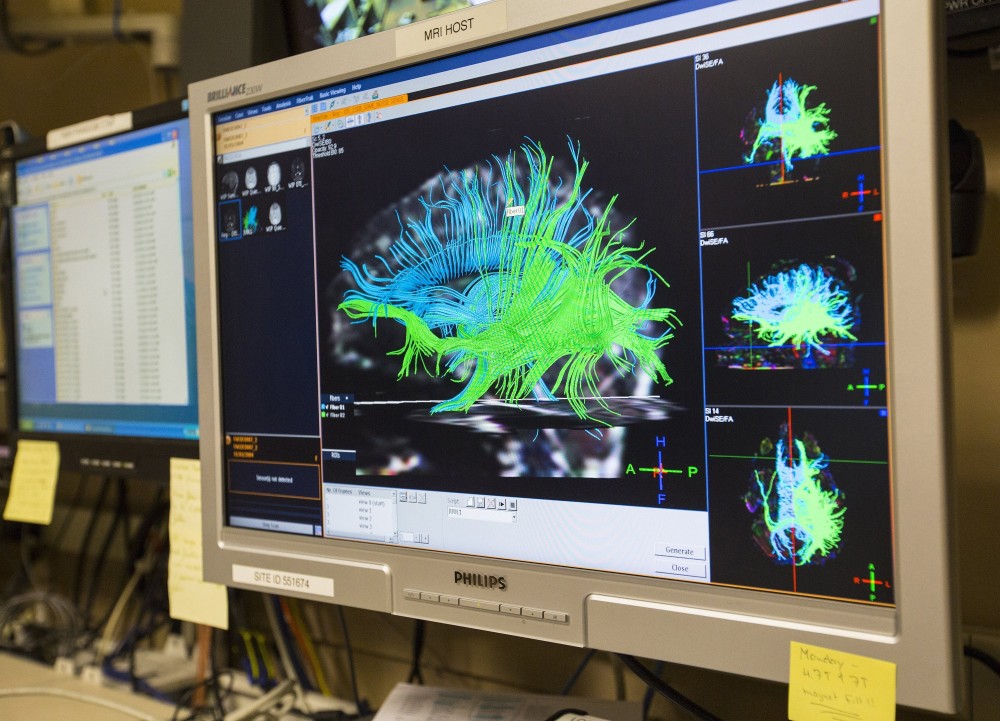
Using a technique called dynamic contrast MRI, scientists could observe glymphatic function in real time. A highlighting agent allowed them to see waste transport in anesthetized animals positioned in each posture.
“It is interesting that the lateral sleep position is already the most popular in humans and most animals, even in the wild,” said Maiken Nedergaard of the University of Rochester, who coined the word glymphatic. Such commonality across species suggests a powerful evolutionary basis for sleeping laterally, she said.
The team additionally proposed that two Alzheimer’s-associated proteins, amyloid-beta and tau, are also subject to disposal via the glymphatic pathway.
Side-sleeping animals flushed away more amyloid and tau than those slumbering on their stomachs or backs. Sticky amyloid causes damaging Alzheimer’s plaques; toxic tau drives the development of neurofibrillary tangles. Both can contribute to mass disruption of brain circuitry.
Benveniste, meanwhile, is launching another brain-waste clearance analysis, this time involving human volunteers.
Back at Cold Spring Harbor Lab, Mitra’s neuronal connectivity work involves the power of physics and mathematics to demystify the brain. He said there are some neurons that have been theorized but never seen.
One of his colleagues at the lab recently uncovered a population of once-elusive neurons and in so doing elucidated how the mammalian organ deals with the element of surprise.
Neuroscientist Adam Kepecs is the first to find brain cells dedicated to sending and receiving messages when something unexpected, good or bad, is perceived.
“It can be any kind of surprise,” Kepecs said.
Initially, there were only indirect lines of evidence suggesting the neurons existed. “They were difficult to isolate,” Kepecs said, “and before certain genetic techniques were available you couldn’t tell which neurons you were recording.”
Recent technological innovations, however, allowed him to see brain responses in real time. Like Benveniste’s analyses, Kepecs’ were conducted only in test animals because it would be unethical to try the procedure in humans.
Kepecs observed the elusive cells via optogenetics, a technique that allows scientists to see genetically altered, light-sensitive neurons in a living rodent’s brain.
The bad surprise was a puff of air in the face; the good one, unexpected food.
He and his colleagues could see “the lights of surprise”, impulses coursing instantaneously through the forebrain of each startled animal. The impulse took only a few thousandths of a second, but it boosted scientific understanding of where surprises are processed. The same cells are likely activated in humans.
“We think that we will be able to map out these neurons … and how they fire and when they broadcast to the rest of the brain,” Kepecs said.
He noted that surprises are processed by “cholinergic neurons,” which means these neurons use the chemical acetylcholine to relay messages. Neurons communicate via electrical or chemical signals. Cholinergic neurons are key in learning and memory, and they are damaged in certain medical conditions.
“These neurons are hit very early in Alzheimer’s disease,” Kepecs said.
Yet, he isn’t venturing any further hypotheses on what his findings mean regarding the devastating brain disorder.
“We have to be cautious,” Kepecs said. “There isn’t an answer right around the corner.”